Figure 2.
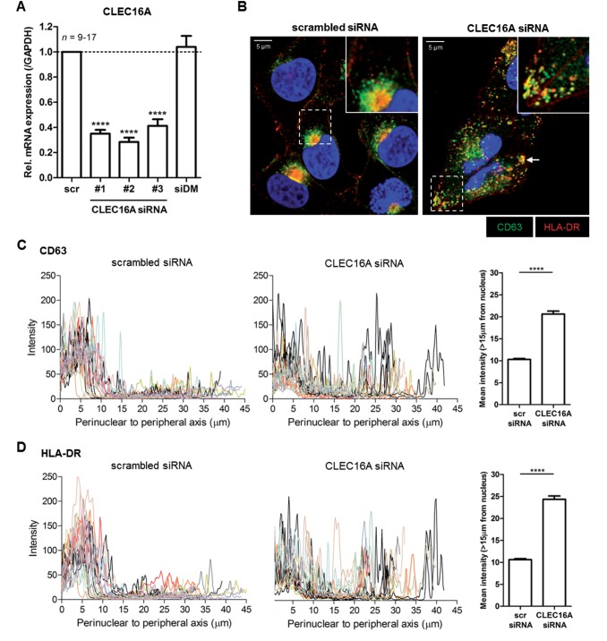
CLEC16A knockdown in antigen-presenting cells causes dispersed localization of HLA-II-positive late endosomes. The human melanoma cell line MelJuSo was transfected with three distinct CLEC16A siRNA duplexes (#1, #2 and #3) and analysed after 3 days. (A) Relative CLEC16A mRNA levels in CLEC16A siRNA- compared to scrambled siRNA-treated cells (n = 9–17). Cells were also transfected with a pool of irrelevant HLA-DMβ siRNA as additional control. Immunofluorescence analysis (B) and quantification of the cytoplasmic localization of CD63+HLA-DR+ late endosomes (C and D) in CLEC16A siRNA- and scrambled siRNA-treated MelJuSo cells (n = 17–21). The images are representative of five experiments. Late endosomal scattering was quantified by analysing fluorescence intensities from the perinuclear area to the outermost peripheral regions (>15 μm from the nucleus; 15 μm cut-off based on scrambled siRNA histograms) for each cell using a line scan (LAS AF software). Scale bars = 5 μm. ****P < 0.0001.

