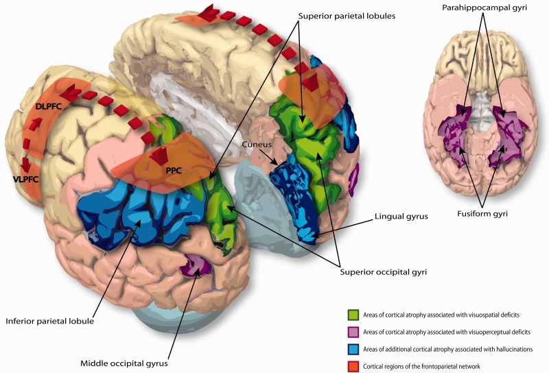
Figure 2.
The major cortical neural networks affected in PDD. Areas of cortical atrophy associated with visuospatial and visuoperceptual deficits in PDD (coloured green and purple, respectively) are based on the data presented in Pereira et al. (2009). Areas of cortical atrophy specifically associated with the presence of visual hallucinations in PDD (coloured blue) are based on the data presented in Goldman et al. (2014a). Functional cortical regions comprising the fronto-parietal attention network (highlighted red) are based on the data presented in Williams-Gray et al. (2008). Cortical regions are identified according to the Allen Brain Atlas for the human brain, and manually drawn onto the corresponding 3D brain image. In this representation the same cortical regions are affected symmetrically in both hemispheres, however in the original studies above the extent of atrophy in these regions was not symmetrical between hemispheres, and varied between individual patients. In the inferior view of the cortex the cerebellum has been removed to expose the fusiform gyri more clearly. DLPFC = dorsolateral prefrontal cortex; PPC = posterior parietal cortex; VLPFC = ventrolateral prefrontal cortex.

