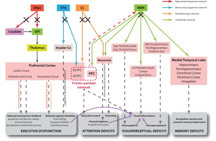
Figure 3.
Hypothetical model of the neural networks affected in PDD and corresponding cognitive deficits. Solid arrows correspond to direct neural connections and colours are indicative of the primary neurotransmitter involved as shown in the key. Dashed arrows connect the relevant dysfunctional neural network to its putative cognitive effects. Purple arrows indicate that a deficit in one cognitive domain contributes to the development of impairment in another domain. Black crosses indicate damage to a neural pathway. The red dashed arrow represents direct projections from prefrontal cortex to the NBM, permitting top-down control of attention from the fronto-parietal network via recruitment of this latter structure and its cortical projections. The limbic, orbitofrontal and associative circuits in the prefrontal cortex correspond to the dissociable fronto-striatal loops of Alexander et al. (1986). Note effects of levodopa therapy at improving and worsening executive functions reliant on cognitive flexibility and learning from feedback, respectively. Electrocortical activation refers to cortical EEG desynchonization indicative of the awake/alert state as described in the text, and is driven by corticopetal cholinergic input from the NBM only. Both cholinergic input from NBM and noradrenergic input from the locus ceruleus (LC) modulate processing in sensory cortices to facilitate orienting of attention to stimuli. Cx = cortex; DLPFC = dorsolateral prefrontal cortex; fx = function; GPi = globus pallidus (internus); PPC = posterior parietal cortex; SNpc = substantia nigra pars compacta; VLPFC = ventrolateral prefrontal cortex; VTA = ventral tegmental area.

