Figure 1.
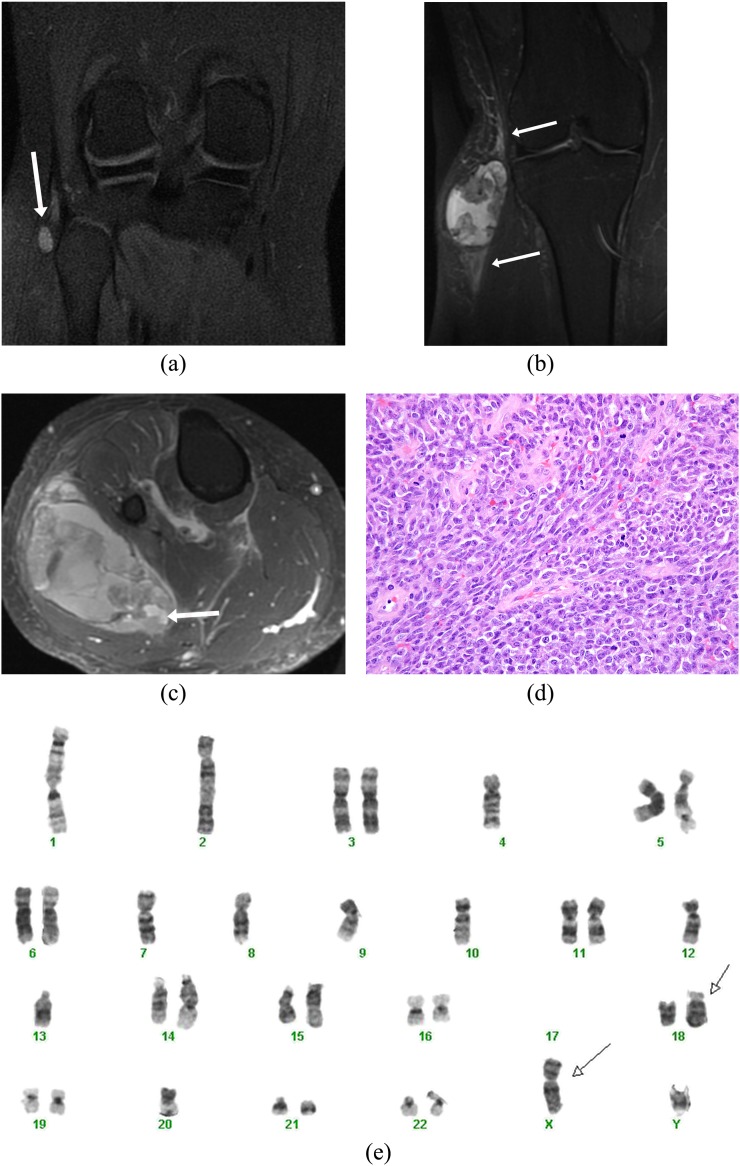
A 21-year-old male presented with a painless “pea-sized” lump on the right knee. (a) Coronal non-contrast inversion recovery sequence image shows an incidental small well-defined hyperintense lesion on the lateral aspect of the knee detected during an MRI knee study (arrow). A contrast-enhanced study was not performed, and the lesion was presumed to possibly represent a ganglion cyst/collection, and follow-up imaging was suggested. (b) The patient was unfortunately lost to follow up and presented 3 years later with a large swelling involving the right knee. Coronal inversion recovery sequence image reveals dramatic increase in the heterogeneous lesion showing hyper-, iso- and hypointense components (triple sign) along with perilesional oedema (arrows). (c) Axial post-contrast fat-saturated image demonstrates heterogeneous enhancement with presence of fluid–fluid levels (arrow) consistent with blood products. The patient unfortunately also had pleuropulmonary metastases and is currently on chemotherapy. (d) Histology showed a poorly differentiated synovial sarcoma composed of spindled to round cells with a high mitotic rate (original magnification, ×400). (e) Cytogenetic analysis showed a complex karyotype (following neoadjuvant therapy), which included the pathognomonic translocation t(X;18)(p11;q11) (arrows).

