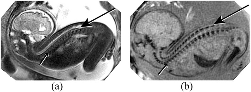
Figure 2.

(a) Sagittal half Fourier acquisition single-shot turbo-spin echo (HASTE) image of a foetus with severe cervical retroflexion. The spinal cord (long arrow) is easily seen surrounded by high signal cerebrospinal fluid. The vertebral column is poorly demonstrated on HASTE imaging (short arrow). (b) Similar plane on susceptibility weighted imaging (SWI). The vertebral column and skull base are well demonstrated, including the laminae (long arrow) and vertebral bodies (short arrow). The spinal musculature is higher signal on SWI than on HASTE, providing significantly improved contrast.
