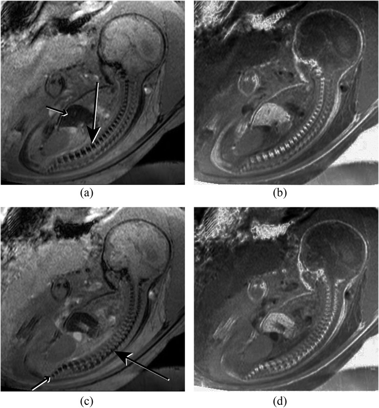
Figure 3.
(a) Sagittal susceptibility weighted imaging (SWI) of the spine nicely demonstrating the vertebral bodies (long arrow). The liver (short arrow) is also low signal owing to the presence of iron. (b) Inverted image of (a) demonstrates an appearance similar to a standard radiograph. (c) Slightly parasagittal SWI image of the spine demonstrating the laminae of the posterior elements (long arrow). The vertebral bodies of the sacrum are also shown (short arrow). (d) Inverted image of (c).

