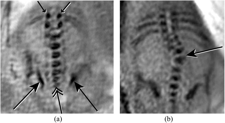
Figure 5.
(a) Coronal susceptibility weighted imaging (SWI) image of the lumbosacral spine demonstrating normal iliac bones (long arrows), lumbosacral vertebral bodies (double arrow) and laminae (short arrows). (b) Coronal SWI image of the spine demonstrating a scoliosis secondary to a vertebral segmentation defect (long arrow).

