Figure 6.
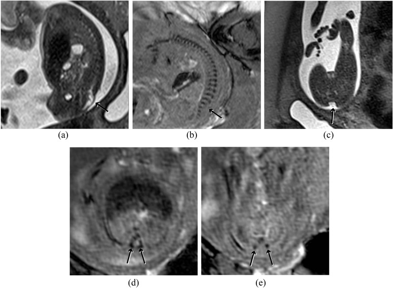
(a) Sagittal half Fourier acquisition single-shot turbo-spin echo (HASTE) image of the spine demonstrating a lumbosacral myelomeningocele (short arrow). (b) Corresponding susceptibility weighted imaging (SWI) image demonstrating the laminae throughout the spine except at the level of the defect (arrow), which can be seen to be at L5. (c) Axial HASTE image demonstrating the dysraphism (arrow), but bony anatomy is poorly demonstrated. (d) Axial SWI image above the lesion demonstrates the laminae pointing towards each other (arrows), which is the normal configuration. (e) Axial SWI image at the level of the lesion demonstrates the laminae to be short (arrows), not pointing to each other and displaced laterally.

