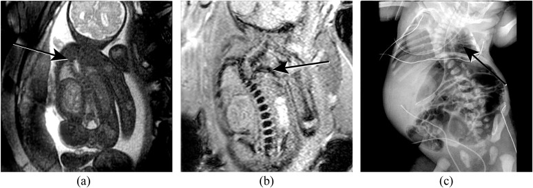
Figure 8.
(a) Coronal half Fourier acquisition single-shot turbo-spin echo view through a foetus with vertebral anomalies, anorectal atresia, cardiac anomalies, tracheo-oesophageal fistula, renal and limb anomalies (VACTERL) association. The thoracic spinal curvature is seen (arrow), but the bony anatomy is not well demonstrated. (b) Coronal susceptibility weighted imaging image better demonstrates the bony anatomy and depicts rib non-segmentation (arrow) at the level of the scoliosis. (c) Postnatal radiograph demonstrates the rib non-segmentation (arrow). Additional rib abnormalities and dextrocardia are also seen.

