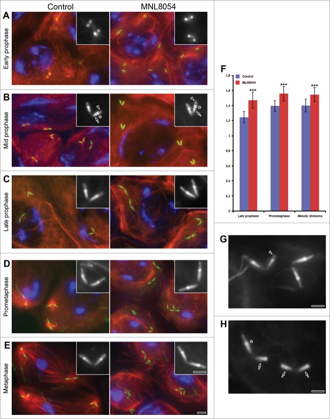
Figure 1.
(See previous page). Aurora-A dependent centriole elongation. Control (left panel) and treated (right panel) primary spermatocytes expressing Unc-GFP were counterstained for acetylated tubulin (red) and DNA (blue). The microtubule cytoskeleton does not show significant differences in control and treated primary prophase spermatocytes (A–C). By contrast, the organization of the cytoplasmic microtubules substantially diverges during prometaphase (D) and metaphase (E) in control and treated spermatocytes: the formers display large asters and bipolar spindles, whereas the others lack both these structures. Magnifications of the centriole/CLR complexes as recognized by Unc-GFP are shown in insets. During early prophase (A) Unc-GFP recognizes small spots in both control and treated spermatocytes. As prophase progressed (B, C) Unc-GFP shows 3 distinct localization: the centriole (arrows, B), the transition region (asterisks, B), the CLR (arrowheads, B). Although the centrioles and the CLRs concurrently elongate during prophase, the centrioles appear slightly longer in late prophase (C), prometaphase (D) and metaphase (E) treated spermatocytes. (F) Quantification of centriole length at different stages of spermatogenesis following 24 hr DMSO (control) or MLN8054 (MLN8054) incubation. Error bars represent SEM. p value from Student's t-test, ***P < 0.0001. Details of abnormally short CLRs (arrowhead, G) or centrioles lacking CLRs (arrows, H); note that the intermediate Unc-GFP dot (asterisk) is only present when the centriole nucleates the ciliary axoneme. Scale bars: (A–E) = 2.5 μm; (A–E) insets, (G, H) = 1 μm.

