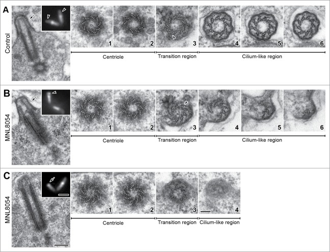
Figure 2.
Defects in CLR organization upon MLN8054 incubation. Longitudinal and cross sections of centriole/CLR complexes in control (A) and treated (B, C) mid prophase primary spermatocytes; insets represent Unc-GFP localization. Control spermatocytes display distinct CLRs evidenced by Unc-GFP (inset A, arrowheads) that protrude from the cell surface (A, arrow). Occasionally, treated spermatocytes display abnormal CLRs (inset B, arrowhead) with tubules of different length (arrow, B) or elongated centrioles that contact the plasma membrane (C), without nucleating the ciliary microtubules (inset C, arrow). The centrioles associated with normal (A1,2) and abnormal (B1,2, C1,2) CLRs display the same architecture; as usual the cartwheel is present in the basal region only. The transition regions display distinct C-blades (A3, B3, arrowheads) when normal (A4–6) or reduced (B4–6) axonemes are present; C-blades are missing (C3) when the axoneme is lacking (C4). Scale bars: (A–C) insets = 1 μm; (A–C) = 250 nm; A1–6, B1–6, C1–4 = 100 nm.

