Figure 2.

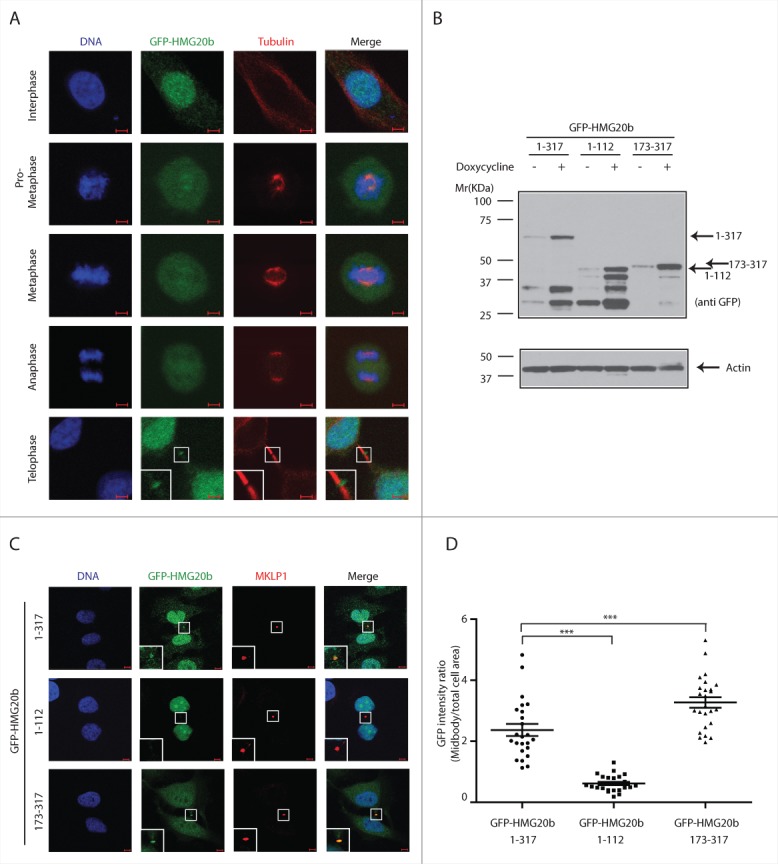
The C-terminal region of HMG20b mediates localization to midbody. (A) Localization of HMG20b during cell cycle. Confocal images of HeLa cells expressing GFP-HMG20b are shown for each cell cycle phase. Enlarged image of midbody is shown as an inset . Scale bar is 5 μm. (B) Western blot showing inducible expression of GFP-HMG20b fragments. β-Actin was probed as a loading control. (C) Confocal images of HeLa cells expressing GFP-HMG20b fragments (green). MKLP1(red) was immunostained as a midbody marker. Enlarged images of midbody are shown as insets. Scale bar is 5 μm. (D) GFP intensity at midbody normalized to the intensity within total cell area for each GFP-HMG20b fragments. A representative scatter dot plot from 3 independent experiments is shown with mean ± s.e.m. Twenty-five cells were analyzed in each experiment. Statistical significance was confirmed for the indicated pair-wise comparisons using Dunnett's multiple comparison test (***: P < 0.001).
