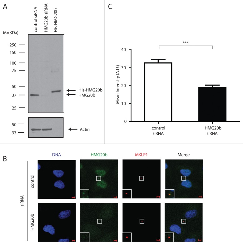
Figure 3.

Endogenous HMG20b localizes to the cytokinetic midbody. (A) Western blot analysis of control and HMG20b siRNA-treated HeLa cell extracts using HMG20b monoclonal antibody 1F6. Purified His-HMG20b protein was used as a positive control and β-Actin was probed as a loading control. (B) Confocal images of control and HMG20b siRNA-treated HeLa cells stained with anti-HMG20b monoclonal antibody 1F6 (green). MKLP1 (red) was co-stained as a midbody marker. Enlarged images of midbody are shown as insets. Scale bar is 5 μm. (C) Mean intensity of HMG20b staining at midbody. Results from 50 cells per sample are shown with mean ± s.e.m. (***: P < 0.001, unpaired t test)
