Figure 5.

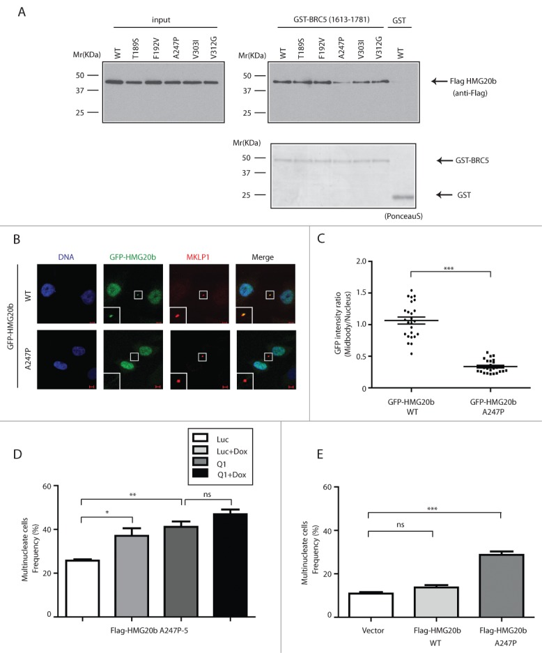
A cancer-associated mutation in the C-terminal region of HMG20b disrupts cytokinesis in a dominant manner. (A) GST-pull down assay. Flag-HMG20b mutants expressed in 293T cells were incubated with GST-BRCA2 fragment containing BRC5. Bound proteins were detected by western blotting using anti Flag antibody. Ponceau S- stained membrane is included in the lower panel to show that equal amount of GST fusion proteins was used for each sample. (B) Confocal images of HeLa cells expressing wild type or A247P mutant of GFP-HMG20b (green). MKLP1 (red) was immunostained as a midbody marker. Enlarged images of midbody are shown as insets. Scale bar is 5 μm. (C) Localization of wild type and A247P mutant of GFP-HMG20b at midbody. A representative scatter dot plot of GFP intensity ratio from 3 independent experiments is shown with mean ± s.e.m. Twenty-five cells were analyzed in each experiment (***: P < 0.001, unpaired t test). (D) Clone (A247P-5) expressing Flag-HMG20b with A247P mutation was analyzed for multinucleation frequency after HMG20b siRNA (Q1) and/or Doxycycline (Dox) treatment. Means ± s.e.m. from 3 experiments are shown and >500 cells were analyzed per each sample. Statistical significance was confirmed for the indicated pairwise comparisons using Bonferroni's multiple comparison test (**: 0.001 < P < 0.01, *: 0.01 < P < 0.05, ns: P > 0.05). (E) Multinucleation frequency measured at 72 hours after HeLa cells were transiently transfected with Flag-HMG20b constructs . Means ± s.e.m. from 4 independent experiments (2 experiments for the vector control) are shown. Transfected cells were identified by anti-Flag immunostaining and >200 transfected cells were analyzed per each sample. Wild type and A247P mutant of HMG20b are compared with the vector control by Dunnett's multiple comparison test (***: P < 0.001, ns: P > 0.05).
