Figure 1.
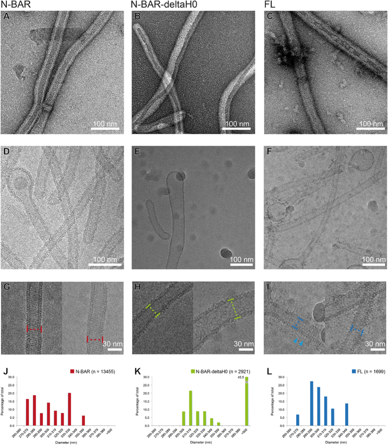
(A–C) Negative-stain EM images of tubes mediated by N-BAR (A), N-BAR-deltaH0 (B) and FL amphiphysin/BIN1(C). (D–F) Cryo-EM images of tubes mediated by N-BAR (D), N-BAR-deltaH0 (E) and FL (F). (G–I) Zoom-in views of N-BAR tubes (G), a thin N-BAR-deltaH0 tube (H, left) and a thick N-BAR-deltaH0 tube (H, right), and FL tubes (I). Markers show the definitions of the width at the outer membrane leaflet. In (I), needle-like densities around tubes are shown (arrowhead). (J–L) Histograms of the distributions of the width of the tubes mediated by N-BAR (J), N-BAR-deltaH0 (K) and FL (L).

