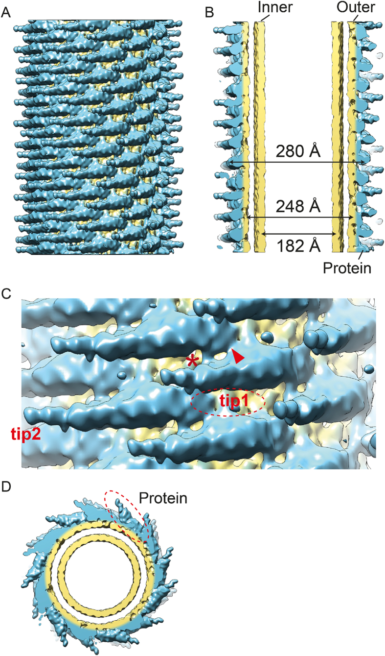
Figure 4.
(A) 3D reconstruction of an amphiphysin/BIN1-mediated tube with a diameter of 280 Å. The density corresponding to the protein is colored in blue and the lipid corresponding parts are colored in in yellow. (B) The central portion of the 3D reconstruction is shown in (A). Inner leaflet (diameter of 182 Å) and outer leaflet (diameter of 248 Å) are colored in yellow. (C) Zoom in view of A. “*” indicates a rod-like density connecting adjacent BAR domains. The red arrowhead indicates an additional density connecting the adjacent BAR domains. “Tip1” shows a tip density hidden under the membrane. “Tip2” shows the second tip of the BAR unit. (D) End-on view of (A).

