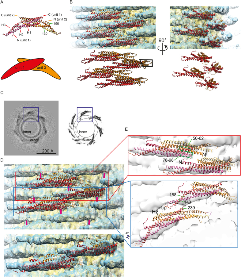
Figure 5.
(A) Atomic model (PDB code 1URU) of Drosophila amphiphysin (top). The crescent BAR unit is achieved by dimerization (unit 1 and unit 2, bottom). The amphipathic H0 helix is not visualized, as it is unstructured in the crystallization condition. (B) Fitting of the atomic model shown in (A) to the amphiphysin mediated tube of 280 Å –diameter. The black box highlights the tip portion not visualized by the reconstruction. (C) The density of a section of the 3D reconstruction (left) in comparison to the densities of the amphiphysin atomic model fitted and symmetrized according to the arrangement calculated from the 3D reconstruction. Inner/outer: headgroup density of the inner or outer leaflet. One tip of the BAR is immersed into the acyl chain region according to the rigid body fitting. This part of the reconstruction was not resolved due to the surrounding lipids (boxed in (B)). (D) (Top) The rod-like density connecting the BAR units are marked with a pink bar. (Bottom) A cropped representation of (top) without the pink marker. (E) Representation of the atomic models showing the arrangement of the neighboring BAR units.

