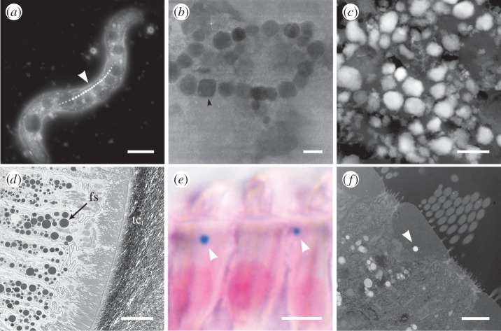
Figure 4.
A selection of electron and optical micrographs of biogenic iron minerals formed by organisms. (a) Dark-field scanning transmission electron micrograph (DF-STEM) of magnetotactic bacterium Magnetospirillum magnetotacticum clearly showing the chain of nanoparticulate magnetite forming the magnetosome (arrowhead), scale bar, 1 µm. (b) Bright-field TEM (BF-TEM) micrograph of chains of magnetite isolated from ethmoid tissue from the salmon Oncorhynchus nerka (reproduced from refrence [30]), scale bar, 50 nm. (c) DF-STEM micrograph of iron granules extracted from trophocyte cells in the abdominal fat layer of the honeybee Apis mellifera, scale bar, 500 nm. (d) BF-TEM micrograph of the mass accumulation of ferritin siderosomes (fs) in the epithelial tissue surrounding the magnetite mineralized tooth cusp (tc) of the chiton Acanthopleura hirtosa, scale bar, 5 µm. (e) Optical micrograph of a Perls' Prussian blue stained section and (f) a DF-STEM micrograph both showing a single iron-rich cuticulosome (arrowheads) in the cuticular plate of inner ear hair cells from the pigeon Columbia livia, scale bars, 5 µm (e), 2 µm (f). Note: bright regions in DF-STEM images correspond to regions of high mass.

