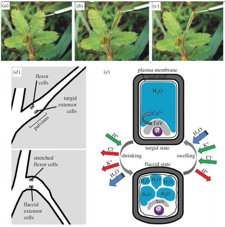
Figure 5.
Thigmonastic movement of leaflets and schematics of mechanisms in Mimosa pudica. (a) Leaflets open; (b) leaflets closing owing to touch-induced changes in cell turgor of cells within the pulvinus, a structure located at the base of each leaflet. (c) Leaflets closed. The time-lapse between each photograph is about 1 s. (a–c) Adapted from [8]. (d) Cartoon illustrates the mechanism of leaf movement in M. pudica before (top panel) and after (bottom panel) application of stimulus. Note the corresponding states of the flexor and extensor cells in each condition, which implies that the extensor cells are the primary effector of pulvinus bending. (e) Cartoon illustrates turgor changes in an extensor cell and the resulting shape changes of the cell as it changes between turgid (above) and flaccid (below) states; here, N denotes the cell nucleus. The primary ionic movements underlying these turgor changes are shown: when the leaves are mechanically simulated, adenosine triphosphate (ATP) dephosphorylating H+ pumps (green) allow for the rapid outward flux of K+ and Cl– ions, which in turn trigger outward osmotic movement of water. Tannin-rich vacuoles (TnV) store Ca2+ to regulate K+ flux. (d,e) Adapted from [8,22]. (Online version in colour.)

