
Figure 12.
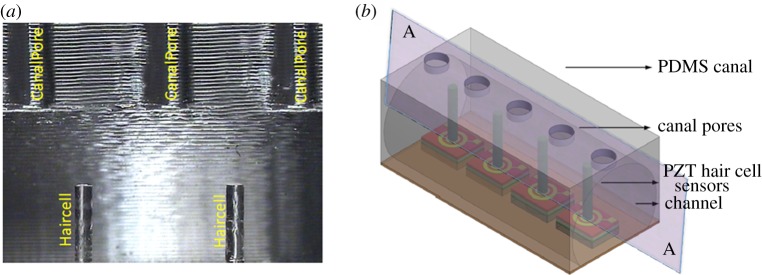
Biomimetic artificial CN sensors. (a) A cross-sectional optical microscopic view of the canal. The canal is mounted in such a way that each hair cell is positioned at the centre of two consecutive pores which lead external flows into the canal. (b) Schematic array of sensors showing the dicing plane along which the microscopic image in (a) is taken. (Online version in colour.)