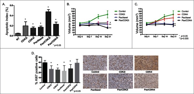
Figure 3.
Paclitaxel/CDK2i induced apoptosis in vitro and decreased tumor volume and Ki67 staining in vivo in a MDA-MB-231 xenograft model. (A) MDA-MB-231 cells were treated and analyzed for apoptosis using TUNEL staining. (B) Female athymic nu/nu mice were inoculated with MDA-MB-231 cells to establish tumors, and treated for 21 d Tumor volume was measured at indicated time points. The tumor volumes from treated groups were compared with the volume from the respective control treated group at Day 21, and significance was then determined; *P < 0.05, **P < 0.005. (C) Representative images from Ki67 stained xenografts from each treatment group at 20X magnification.

