
Figure 1.
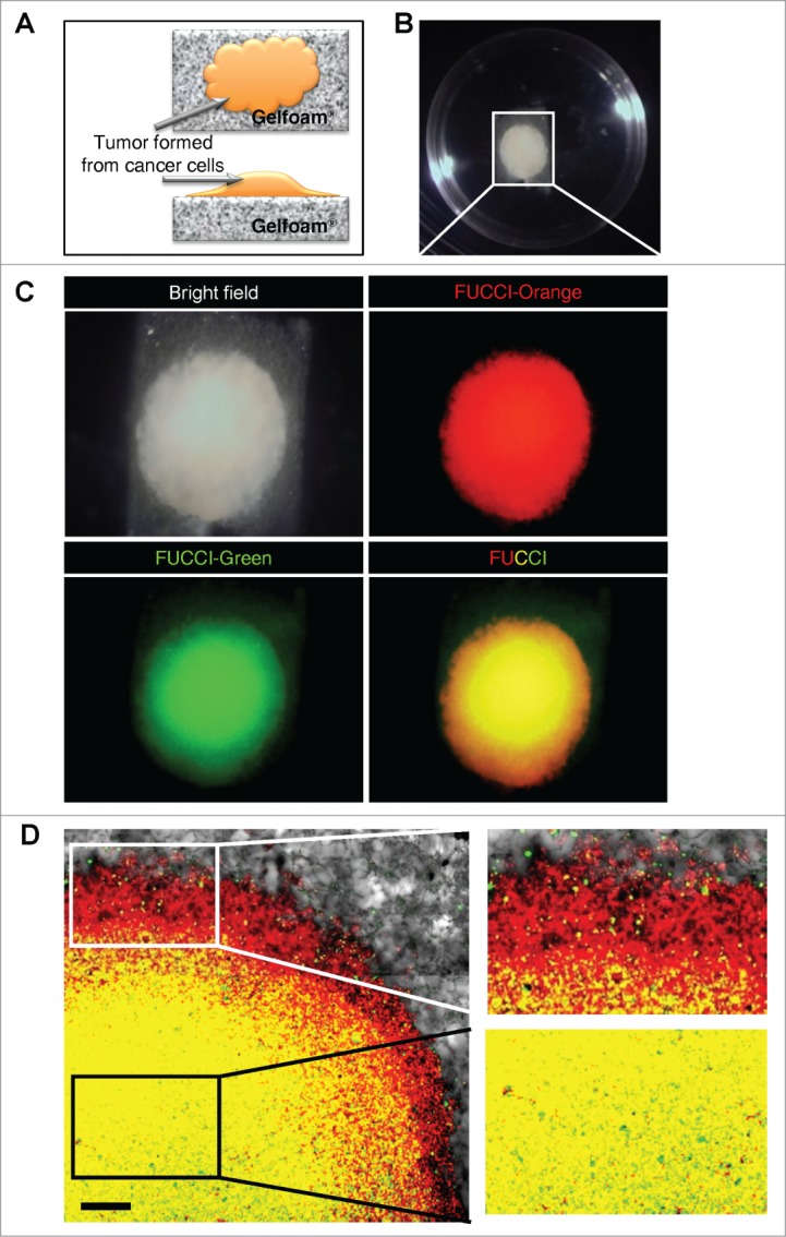
Gelfoam® histoculture of FUCCI-expressing cancer cells. (A) Schema of FUCCI-expressing MKN45 stomach cancer cells forming a tumor on Gelfoam®. (B) Macroscopic appearance of the tumor formed on Gelfoam® histoculture. (C) Macro images of a tumor formed on Gelfoam® demonstrating FUCCI fluorescence. (D) FUCCI-expressing cancer cells in the tumor formed on Gelfoam®. Images at the single-cell level were acquired by confocal laser-scanning microscopy. High magnification images (×10) of an invading area of the tumor (upper right) and a non-invading area (lower right) of the tumor on Gelfoam®.