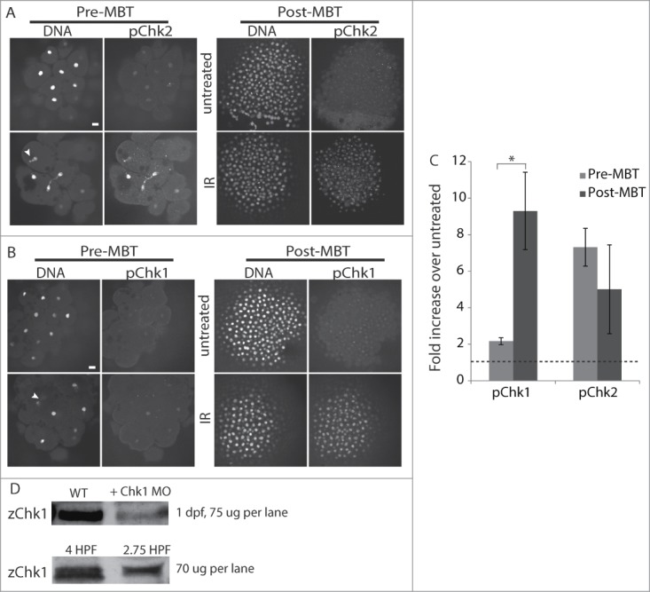
Figure 5.

Pre-MBT embryos can activate Chk2 but not Chk1. (A–C) Pre-MBT (1.5 HPF) or post-MBT (4 HPF) embryos were treated with 10 Gy IR as indicated, then fixed and stained for pChk2 (A) or pChk1 (B) and DNA. Arrowheads show examples of nuclear fragmentation and lagging chromosomes from DNA damage. pChk1 and pChk2 staining were quantified, averaged over multiple embryos (n ≥ 8, pooled from multiple experiments), and plotted (C) as the fold increase of IR treated over untreated. The dashed line represents no increase over untreated control. Error bars indicate SD, *P < 0.05, scale bars 20 μm. (D) Western blots for Chk1 using zebrafish-specific anti-Chk1 antibody. Top blot: zChk1 polyclonal antibody was validated first in embryos 1 day post fertilization. Lanes show 75 ug of total protein from control embryos and 75 ug of total protein from embryos injected with a Chk1 morpholino to confirm the correct band. Bottom blot: 70 ug of total protein was loaded into each lane for 4 and 2.75 HPF embryos.
