
Figure 1.
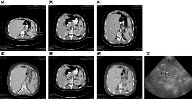
Axial contrast-enhanced CT images through upper abdomen show multiple hypodense lesions (A, B, C) in the spleen (S) consistent with splenic abscesses, associated with mild splenomegaly. A follow-up CT scan performed on the seventh day of admission showed a volumetric reduction of the three abscesses (D, E, F). A follow-up ultrasound images through upper abdomen (G) shows a normal splenic tissue. The tissue is homogeneous in texture and the margins of the capsule (black arrows) are smooth.