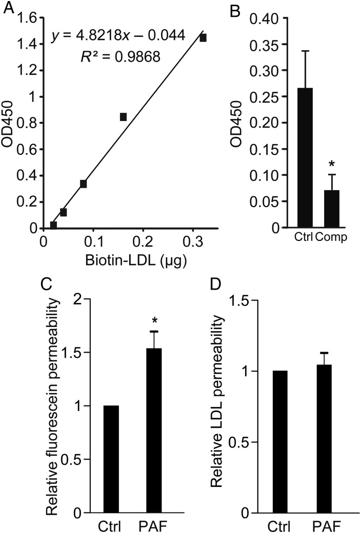
Figure 3.
LDL transcytosis is performed by HCAECs in vitro. (A) Standard curve for ELISA for biotin-LDL showing linearity and high sensitivity; y-axis is optical density at 450 nm (see Methods). (B) Transwells coated with confluent HCAECs were allowed to bind biotin-LDL and then rinsed to remove unbound ligand. Two hours later, LDL in the lower chamber of the transwell was measured by ELISA. Addition of 50-fold excess unlabelled LDL (Comp) reduced biotin-LDL transcytosis, *P < 0.05 by Student's t-test. n = 7 experiments. (C and D) The transwell assay for LDL transcytosis is not affected by paracellular leak. Administration of PAF (10 µM) increased the permeability of endothelial monolayers to Na-fluorescein (C) but did not affect LDL transcytosis (D) compared with untreated cells (Ctrl); *P < 0.05 by one-sample t-test, n = 6 experiments.

