Figure 4.
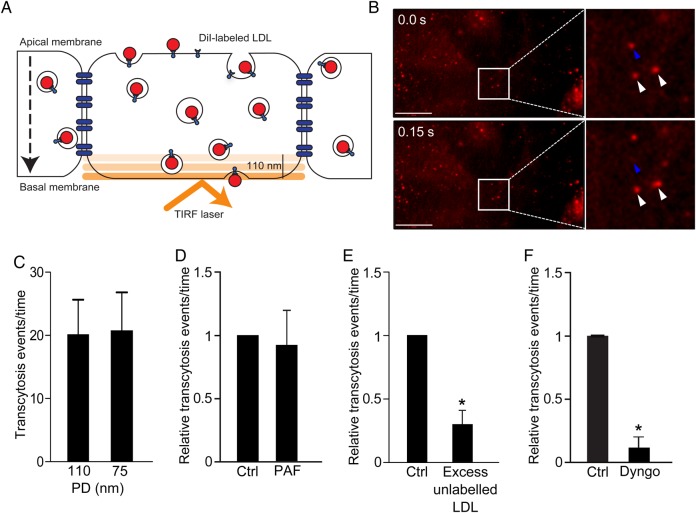
Novel assay to study LDL transcytosis. (A) Schematic of the assay. DiI-LDL is added to confluent endothelial monolayers and is detected by TIRF microscopy as it enters the bottom of the cell. (B) Still images from the Supplemental Video. Top panel depicts initial image, bottom panel is 150 ms later. Blue arrowhead denotes LDL-containing vesicle that disappears due to exocytosis; white arrowheads show vesicles that do not fuse. (C) As a control for endocytic traffic, the penetration depth (PD) of the TIRF laser was decreased and exocytic events were quantified. Changing the PD of the TIRF laser had no effect; n = 4. (D) The TIRF assay is not affected by paracellular leak. Endothelial monolayers were treated with PAF as in Figure 2, and transcytosis events were quantified; there was no significant difference between control (ctrl) and PAF-treated cells, n = 5. (E) Addition of 50-fold excess unlabelled LDL at the same time as DiI-LDL reduced transcytosis, *P < 0.01 by one-sample t-test, n = 4. (F) Pre-incubation for 30 min with Dyngo4A (30 µM), a specific inhibitor of the GTPase dynamin, blocked LDL transcytosis; *P = 0.001 by one-sample t-test, n = 4 experiments.

