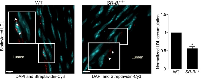
Figure 7.

Deficiency of SR-BI reduces LDL transcytosis ex vivo. Aortas from SR-BI-deficient mice and wild-type (WT) littermates were perfused ex vivo with biotinylated LDL (25 µg/mL). Serial sections were stained with streptavidin-Cy3, and z-stack images were then acquired by confocal microscopy under identical microscope settings. Nuclei are stained with DAPI (blue). All perfusion, sectioning, staining, and image analysis were performed as four separate experiments, each containing one WT and one SR-BI−/− mouse. Images were quantified using ImageJ for Cy3 (red) fluorescence; all signal above background was measured. For each experiment, the same background correction was applied to images from both the knockout and matching wild-type aorta. Results are presented as the red fluorescence intensity in a SR-BI−/− aorta normalized to its WT equivalent. More punctae of LDL are visible in WT vs. SR-BI−/− vessels (inset, arrowheads). Quantification is shown at right. *P < 0.01 by one-sample t-test, four mice per group.
