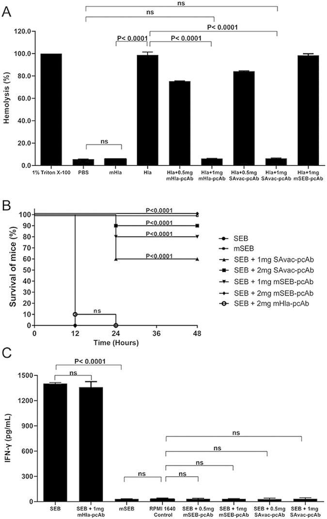
Figure 6. pcAb inhibits the biological function of Hla and SEB in vitro.

(A) Hemolytic activity assay, 1% rabbit erythrocytes diluted with PBS were incubated at 37 °C for 30 min with mHla, Hla, or Hla pre-incubated with mHla-pcAb or SAvac-pcAb, as indicated. The supernatant was then isolated by centrifugation and the hemolytic activity was determined by the release of hemoglobin, measured spectrophotometrically at 540 nm and presented as % hemolysis of the positive control (Triton X-100). Experiments were conducted in triplicate and data were presented as mean values ± standard deviation (SD). (B) Intestinal toxin activity assay, mice were injected i.p. with D-Galactosamine (200 mg/mL) followed by an intramuscular injection with the indicated concentrations of SEB, mSEB, PBS, or SEB incubated with mSEB-pcAb and SAvac-pcAb for 30 min prior to the start of the study. Survival was monitored for 40 h after the challenge and mHla-pcAb was used as a control, the differences between each group were presented as p-value. (C) IFN-γ production assay. Pig splenic leukocytes were isolated and cultured in the presence of the indicated concentrations of wild type SEB or mSEB. After 40 h of culture, supernatants were taken and ELISA was used to determine IFN-γ secretion. mHla-pcAb was used as control and data were presented as mean values ± standard deviation (SD). Experiments were conducted in triplicate.
