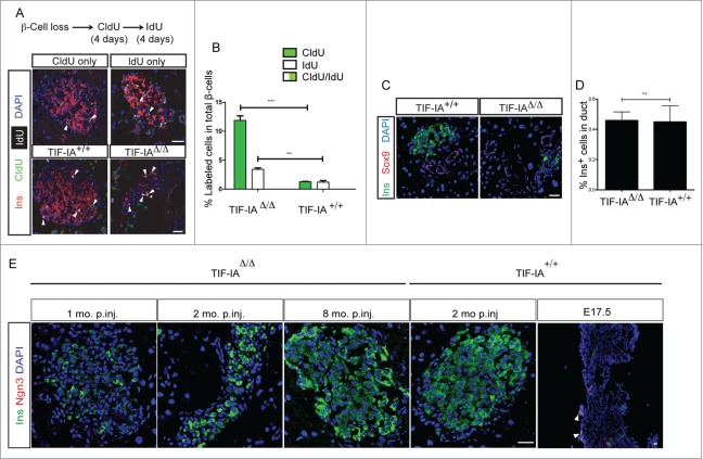
Figure 5.
The new β-cells do not arise from a rapidly proliferating progenitor cell type. (A and B) double thymidine analog labeling shows even distribution of proliferation in the β-cell population. (C and D) The frequency of insulin positive cells inside the duct structures, marked by Sox9 expression, does not change in response to β-cell loss in TIF-IAΔ/Δ mice. E: Ngn3 expression is not induced at several time points after tamoxifen injection. Scale bar: 25 μm. Values are presented by mean ± SEM. P-value <0.001: *** , P-value <0.0001: ****. N > 3 animals for each experimental condition.

