
Figure 2.
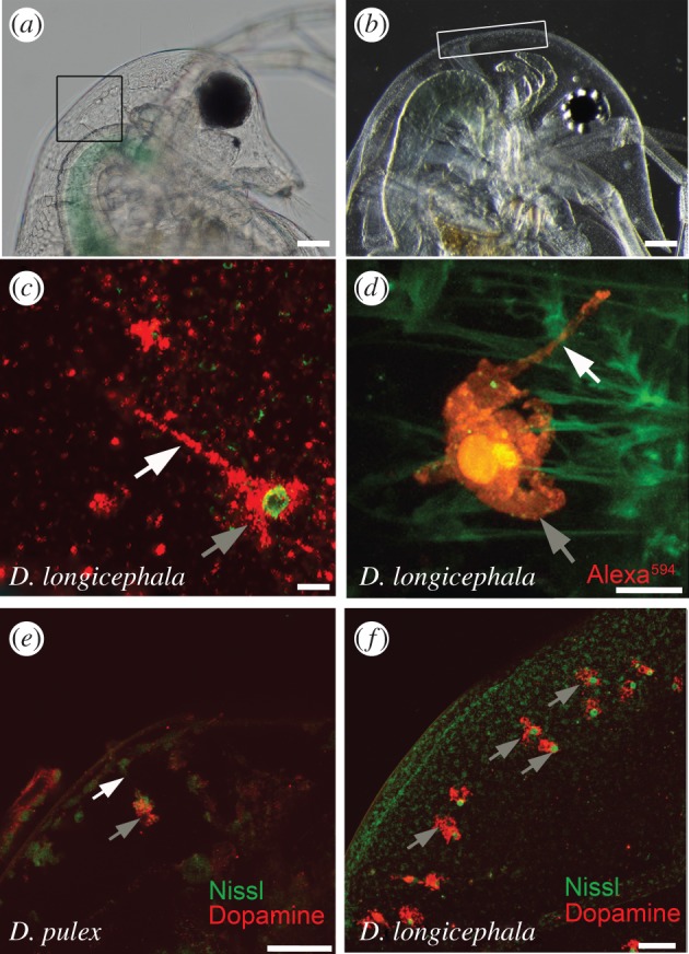
Intracellular localization of dopamine. (a) Daphnia pulex head; box depicts the area of dopamine-containing polyploidy cells. (b) Daphnia longicephala head; box depicts the area of dopamine-containing polyploidy cells. (c) Bulged cell in Notonecta-exposed D. longicephala showing the soma with the nucleus and the cellular extension containing dopamine. Scale bar, 10 µm. (d) Overview of an Alexa594-filled bulged cell with soma and cellular extension. Scale bar, 10 µm. (e) Dopamine immunolabelling of bulged cells in predator-exposed D. pulex. Scale bar, 50 µm. (f) Dopaminergic bulged cells in predator-exposed D. longicephala. Scale bar, 50 µm. (c–f) Grey arrows point to soma of bulged cells, white arrows indicate the cellular extension of the cell.