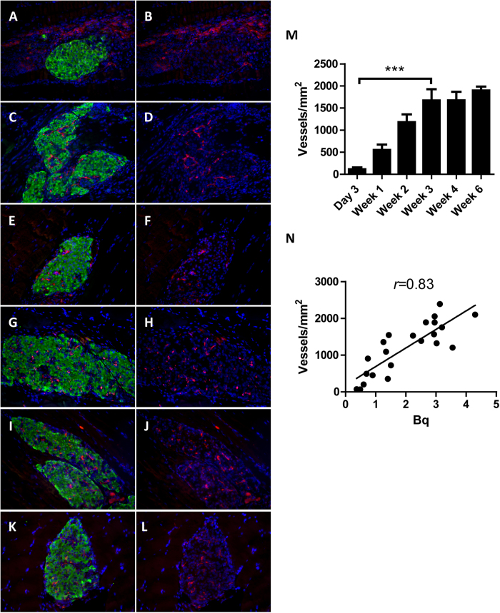
Figure 4. Histological analysis of islets revascularization in the skeletal muscle.
(A–L) Representative images of 3 days to 6 week-old islet transplants for which β-cells were stained with anti-insulin (green) and the ingrowing VEGFR2-expressing capillaries (red). All sections were counter-stained with DAPI (blue). (M) Quantification of vascular density within the insulin positive area shows significant increase in number of vessels from 3 days to 3 weeks (***p < 0.001). N: Initiation and increase of islets vascular density correlates with increasing exendin-3 uptake (pearson r = 0.83). Data are shown as means ± SEM from 4–5 animals.

