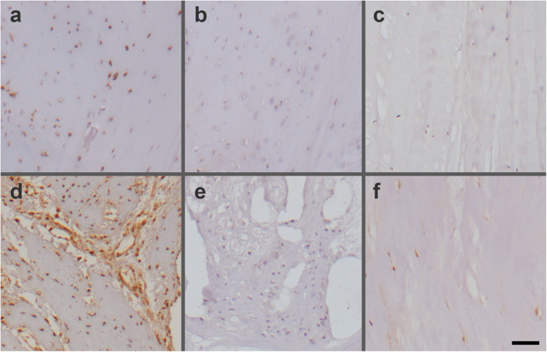
Figure 7.
Immunohistochemical analysis of IGFBP3 (a–c) and IFIT3 (d–f) in sections of human degenerative (a, b, d, e) and non-degenerative (c, f) annulus fibrosus. Intense immunolabelling for IGFBP3 (a) and IFIT3 (d) was observed in degenerated regions of the annulus fibrosus of a lumbar disc from a 47 year old female with disc degeneration Grade III. Negative control sections for IGFBP3 (b) and IFIT3 (e) did not show any staining. IGFBP3 positive cells were absent (c), while IFIT3 positive cells were observed in the inner annulus fibrosus of a normal disc (f) (degeneration Grade I) from a 13 year old male. Scale bar: 50 μm.

