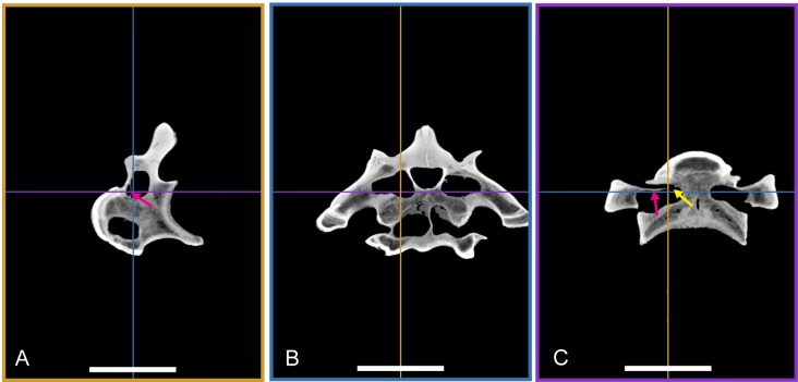
Figure 16. Multi planar reformat (MPR) image of BSPG 2011 I 115.
(A) slice through right lateral aspect of vertebra; (B) transversal slice through vertebra in posterior view, (C) horizontal slice through centrum. All sections are orthogonal to one another, with colors indicating relations between the images. The yellow arrow shows connection of posterior and anterior pneumatic cavities of transverse process; The pink arrow shows thin lamina separating cavities laterally. Scale bars equal 10 cm.

