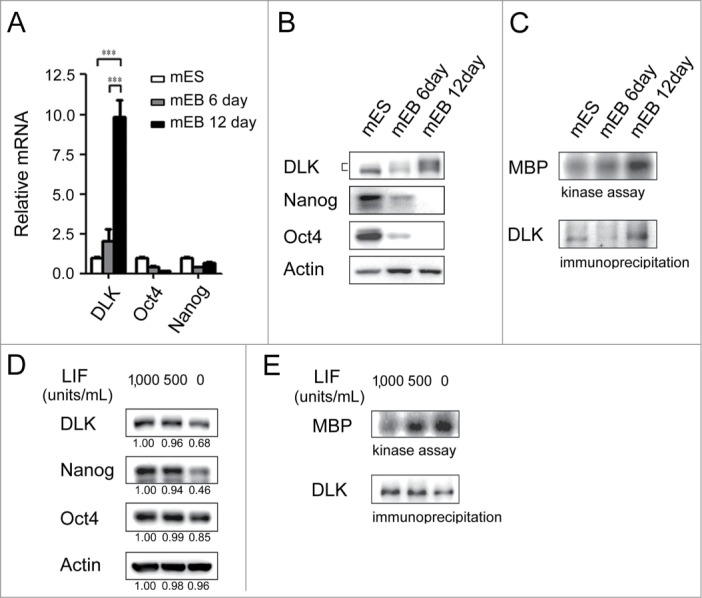
Figure 2.

DLK activity is upregulated upon differentiation. (A) The mRNA expression levels of DLK increased upon differentiation. DLK, Nanog, and Oct4 transcripts from D3 mouse ES cells and mouse EBs (6th and 12th days) were quantified by real-time quantitative PCR analyses. Mouse EBs were prepared by suspension method described in the material and methods. (B) The protein expression level of DLK, Nanog, and Oct4 proteins in mouse ES cells and EBs were detected by Western blotting. (C) DLK enzyme activity increased upon differentiation (EB). DLK protein in mouse ES cells and EBs (6th and 12th days) were precipitated with DLK antibody, and aliquoted for DLK kinase activity assays and Western blotting. Myelin Basic Protein (MBP) was used as a DLK substrate to evaluate DLK kinase activity. (D) Nanog protein level decreased upon the removal of LIF. Mouse ES cells maintained in feeder-free conditions were cultured with medium contained 1,000 units/ml of LIF, 500 units/ml of LIF, or without LIF. These cells were harvested for analysis of DLK, Nanog, and Oct4 protein expression by Western blotting. (E) DLK kinase activity increased upon LIF removal or reduction. Mouse ES cells maintained in feeder free conditions were cultured with medium contained 1,000 units/ml of LIF, 500 units/ml of LIF, or without LIF. DLK kinase activity was measured. The error bars in the figures represent standard error of the mean (mean±SEM). P values were obtained from 2-tailed Student's t-tests (***, P < 0.0001; **, P < 0.001; *, P < 0.05).
