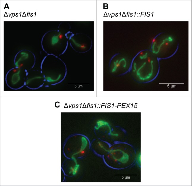
Figure 1.

Peroxisome and mitochondrial fission defects in various yeast mutant strains. Fluorescence microscopy images showing mitochondrial and peroxisome morphology in Δvps1Δfis1 (A), Δvps1Δfis1::FIS1 (B) and Δvps1Δfis1::FIS1-PEX15 (C) cells. Cells were grown until the mid-exponential growth phase on MM containing 2% glucose. Peroxisomes are marked by DsRED-SKL and mitochondria by mitoGFP.
