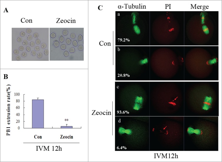
Figure 2.

Severe DNA DSBs oocytes did not reach the MII stage at 12 h of IVM. (A) Oocytes with or without zeocin treatment were maturated in M2 medium for 12 h. (B) The percentage of PB1 extrusion in oocytes undergoing GVBD at 2 h of IVM was analyzed at 12 h of IVM. Two asterisks indicate dramatically significant difference compared to the control group (P < 0.01). (C) Spindle assembly and chromosome alignment at 12 h of IVM. Oocytes in the 2 groups were stained with anti-α-tubulin-FITC antibody and PtdIns. The percentages of each type are indicated.
