Figure 6.
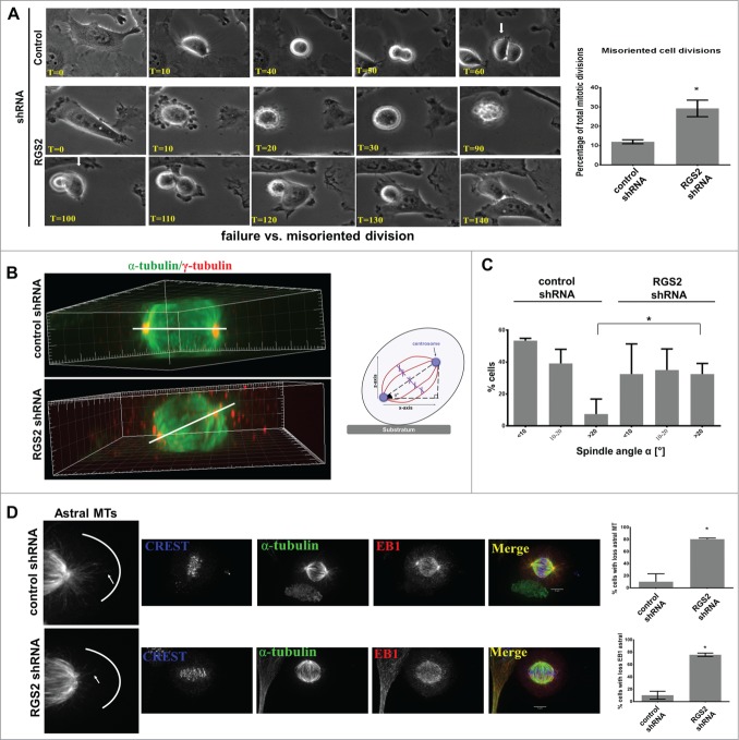
Loss of RGS2 causes spindle misorientation. (A) Frame series of time lapse live imaging movies show incorrect flattening of RGS2-depleted daughter cell onto the substrate after mitosis (misoriented cell division) (see text for details). Also uneven timing of division completion can be observed. Arrows indicate when the first daughter cell begins flatting. T = Time , min. Right: Graphs showing the increased percentage of misoriented cell division in RGS2-depleted cells. *P<0.05. (B) Side view of reconstructed 3-dimensional image shows RGS2-depleted cell exhibiting a high degree of spindle tilting relative to the cell-substratum adhesion plane compared to control cell. Spindle angle (α°) (bar) in RGS2 depleted-cell = 60°; α° in control cell = 0°. Right: Schematic of the angle of rotation in misoriented cell division. (C) The spindle angle is increased in RGS2-depleted cells. Graphs show RGS2-depleted cells with significant increase of spindle angle >20°. Spindle (α-tubulin), centrosomes (γ-tubulin) and/or PLK1 were used to identify spindle poles. Data represent means and standard deviations of a total of 75 mitotic cells in 3 independent experiments. n = 25 counted cells/experiment. *P < 0.05 (D). RGS2 depletion impairs the astral MTs recruitment to the cell cortex. Left enlargements show astral MTs at the cell cortex. Right, graphs show the phenotypes quantification. n = 40 counted cells/triplicated experiments. *P < 0.05.

