Figure 3.
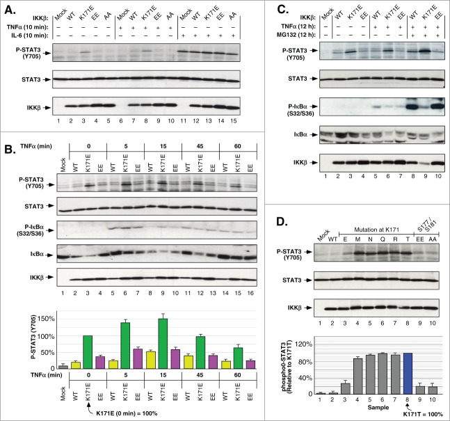
IKKβ Lys171 Activation of STAT3. (A) HEK293T cells expressing IKKβ derivatives were treated with 10 ng/ml TNFα or 10 ng/ml IL-6 for 10 min. Lysates were immunoblotted for Phospho-STAT3 (Tyr705) (top), STAT3 (middle panel) and IKKβ (bottom). (B) HEK293T cells expressing IKKβ mutants were treated with 10 ng/ml of TNFα for 5, 15, 45 and 60 min. Lysates were immunoblotted for Phospho-STAT3 (Tyr705) (top), STAT3 (2nd panel), Phospho-IκBα (Ser32/36) (3rd panel), IκBα (4th panel), and IKKβ (bottom). Phosphorylation of STAT3 was quantitated relative to the K171E mutant at time zero, +/− sem. (bottom panel). (C) HEK293T cells expressing IKKβ WT, K171E and S177E/S181E were treated with +/− 10 ng/ml TNFα and +/− 10 μM MG132 for 12 h. Lysates were immunoblotted as in (B). (D) Lysates from HEK293T cells expressing IKKβ derivatives were immunoblotted as in (A). STAT3 phosphorylation was quantitated relative to the Lys171Thr mutant, +/− sem. (bottom).

