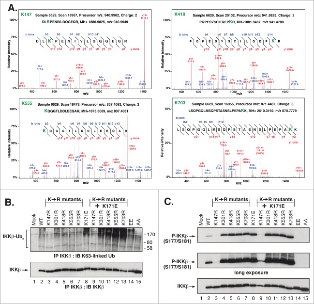
Figure 4.
Identification and Biological Function of IKKβ ubiquitination Sites. (A) Representative spectrum of peptides showing the relative intensity of selected b and y ions for the major sites of IKKβ ubiquitination. Note that not all identified ions are labeled due to space constraints. (B) HEK293T cells expressing IKKβ Lys to Arg mutants were immunoprecipitated and immunoblotted as in Figure 2A. (C) Lysates from HEK293T cells expressing IKKβ Lys to Arg mutants were immunoblotted as in Figure 1(C).

