
Figure 6.
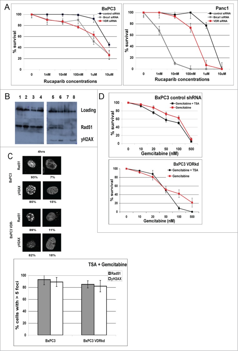
For figure legend, see page 3850.Figure 6 (See previous page). VDR knockdown sensitizes BXPC3 and Panc1 cells to the PARP inhibitor, Rucaparib. blocks gemcitabine induced DSB HR repair by reducing Rad51 foci formation at DSBs. (A) Rucaparib kill curves generated from clonogenic assays of cells transfected with control, BRCA1 and VDR siRNAs. (B) Western blot comparing Rad51 and γH2AX protein levels of BxPC3 and BxPC3 VDRkd cells. 40μg of protein loaded. Lane 1 = BxPC3 + vehicle (18 hrs) (supernatant fraction); Lane 2 = BxPC3 + gemcitabine (50 nM) (18 hrs) (supernatant fraction); Lane 3 = BxPC3 VDRkd + vehicle (18 hrs) (supernatant fraction); Lane 4 = BxPC3 VDRkd + gemcitabine (50 nM) (18 hrs) (supernatant fraction); Lane 5 = BxPC3 + vehicle (18 hrs) (pellet fraction); Lane 6 = BxPC3 + gemcitabine (50 nM) (18 hrs) (pellet fraction); Lane 7 = BxPC3 VDRkd + vehicle (18 hrs) (pellet fraction); Lane 8 = BxPC3 VDRkd + gemcitabine (50 nM) (18 hrs) (pellet fraction). (C) Comparison of Rad51 and γH2AX staining of BxPC3 and BxPC3kd cells following TSA (500 nM) + gemcitabine (50 nM) treatments. (D) Colony survival of BxPC3 VDRkd and BxPC3 parental cells treated with TSA (500 nM) + gemcitabine (n = 3). p values: BxPC3 VDRkd = 0.289, BxPC3 control = 0.02.