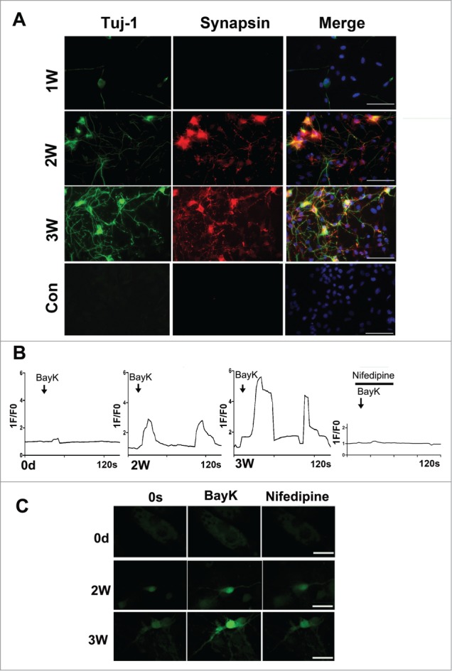
Figure 5.

Characterization of the neuron-like cells derived from MEFs. (A) Immunostaining of the neuron-like cells at 1–3 weeks post-induction with synapsin and Tuj-1 antibodies. scale bar, 50 μm. (B) Functional characterization of the L-type Calcium channel. The panels show typical calcium imagines response observed in neuron-like cells at 2–3 weeks post-induction. 1F/F0 represents the ratio of fluorescence intensity of cells for 0s and an indicated time. Bay-K (10 μM) in the presence or absence of nifedipine (5 μM) was added at the marked time point. (C) The represent figures of calcium imaging of the induced neurons or MEFs with or without nifedipine following in the treatment with BayK. The total number of cells analyzed in each experiment was 40–50 cells, represent results were present. scale bar, 20 μm.
