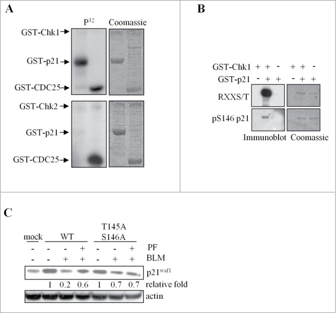
Figure 5.

Chk1 phosphorylates in vitro p21Waf1 at sites involved in p21Waf1 degradation. (A) In vitro kinase assays performed with recombinant GST-Chk1 (upper) or GST-Chk2 (lower) kinases and GST-p21Waf1 or GST-Cdc25C fragment as substrates. Low amounts of kinases were added to increase the specificity of the assay. Left panel: autoradiography of incorporated 32P; right panel: Coomassie staining. (B) The non radioactive in vitro kinase assay containing GST-Chk1 and GST-p21Waf1 proteins was immunoblotted with an anti-RXXpS/T specific antibody or an anti-pS146- p21Waf1 antibody. Normalization was done by Coomassie staining. (C) U2OS cells were transfected with wt or T145A/S146A p21Waf1 double mutant. Tests were conducted 72 hrs after transfection, when the expression levels of the exogenous proteins were reduced to less than fold2- of the endogenous p21Waf1 and to avoid possible effects of the overexpression on cell cycle progression. Cells were pre-treated or not with 400 nM PF-477736 for 1 hr and then exposed to 120 μM BLM for 3 hrs. Whole cell extracts were analyzed by protein gel blot. The densitometric analyses of the experiment, normalized to actin and to mock value, are shown.
