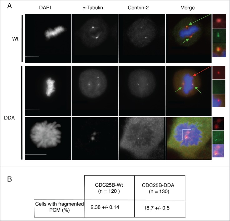
Figure 3.

Multipolar spindles in U2OS-CDC25B-DDA cells are the result of PCM fragmentation. (A). Representative fluorescent microscopy images of asynchronously growing U2OS cells in metaphase or prophase that express CDC25B-Wt or CDC25B-DDA fixed and stained with anti-γ-Tubulin and anti-Centrin-2 antibodies. DNA was stained with DAPI. Centrosomes are highlighted by green arrows, and PCM fragmentation by a red arrow (magnified view in insets). In the merged images, yellow shows co-localization of Centrin-2 and γ-Tubulin. Scale bar: 10 μm. (B). The number of cells with fragmented PCM (i.e., cells with an excess of γ-Tubulin dots) was quantified in U2OS-CDC25B-Wt and U2OS-CDC25B-DDA cells. Values are the mean ± SD of 3 independent experiments.
