
Figure 3.
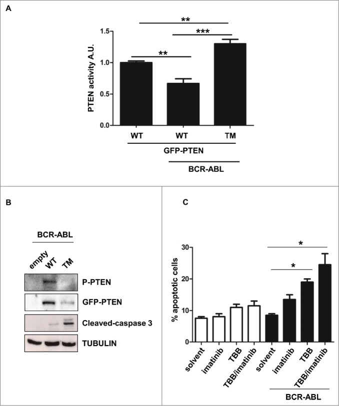
PTEN reactivation promotes apoptosis in BCR-ABL positive cells. (A) Parental and BCR-ABL-NIH3T3 were transfected with the indicated GFP-tagged-PTEN vectors. After 48 hours, PTEN phosphatase assay was performed in immunoprecipitated GFP-PTEN. *P <0.05; **P<0.01. (B) Western immunoblot of BCR-ABL-NIH3T3 cells transfected with the indicated GFP-PTEN mutants. Phospho-PTEN: PTEN-S380/T382/T383. (C) Apoptosis was assessed in NIH3T3 cells treated with 60 μM TBB and 1 μM Imatinib for 10 hours. Data are mean and d.s. of two independent experiments. After short incubation period with inhibitors, in BCR-ABL-NIH3T3 cells, solvent vs. TBB and solvent vs. TBB/imatinib differences are significant with *P<0.05.