
Figure 4.
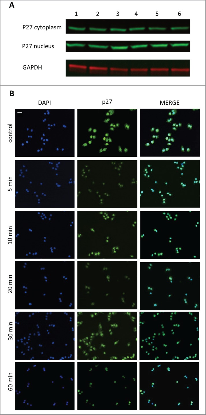
The effect of GHRH antagonist on the localization of p27 in A-375 human malignant melanoma cells determined by Western blot (A) and immunocytochemistry (B). The level of p27 was detected in cytoplasmic and nuclear fractions of A-375 cells with Western blot following incubation with 10 μM MIA-690 for 5, 10, 20, 30 and 60 min (A). Internal standard for the cytoplasmic fraction was GAPDH. Changes in the intracellular localization of p27 following incubation with 10 μM MIA-690 was also detected by immunocytochemistry. In this experiment GHRH antagonist was again added for 5, 10, 20, 30 and 60 min. Immunostained coverslip samples were imaged with a Nikon Eclipse Ti Inverted Microscope fitted with epifluorescence optics and images were recorded using a Nikon DS-Qi1Mc camera. Nuclei are labeled with DAPI (blue). Bars correspond to 100 μM.