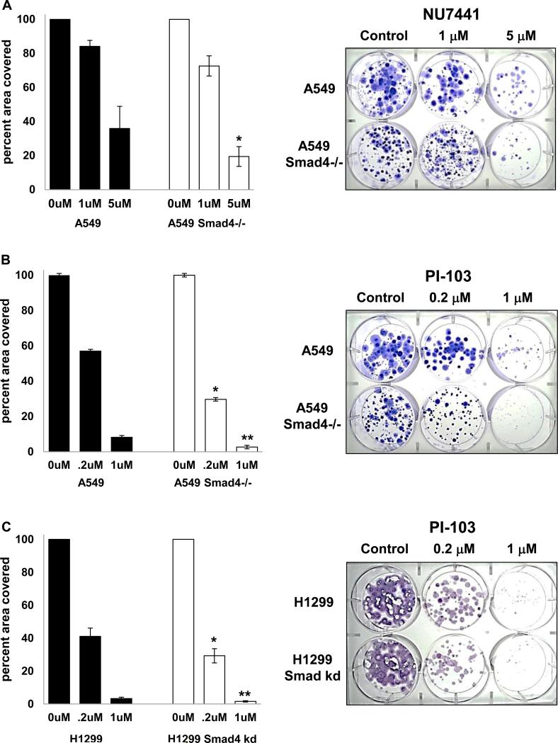
Figure 7. Reduced Smad4 expression increases sensitivity to DNA-PK inhibitors.
(A) A549 Smad4−/− cells were grown in NU7441 at the indicated concentrations for 17-23 days then stained with crystal violet. * p=0.05 vs A549. (B) A549 Smad4−/− cells (100/well) were grown in PI-103 at the indicated concentrations for 12-23 days and then stained with crystal violet. * p=0.02 vs A549; ** p=0.08 vs A549. (C) H1299 cells with Smad4 knockdown (100/well) were grown in PI-103 at the indicated concentrations for 12-17 days and then stained with crystal violet; * p=0.07 vs H1299; ** p=0.02 vs H1299. Examples of long term proliferation assays are shown at right.

