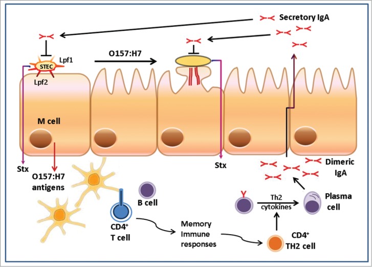
Figure 1.

Working model for EHEC O157 colonization and immune responses required to confer protection. The diagram shows human intestinal epithelia displaying an M cell and the first stages of colonization by Shiga toxin-producing E. coli (STEC) or enterohemorrhagic E. coli (EHEC) O157:H7 where bacteria interact with the intestine via the Lpf fimbriae or other adhesins, followed by the formation of type III secretion-mediated A/E lesion. Luminal O157 antigens are transported through the intestinal Peyer's patches where dendritic cells process and present antigens to T cells. CD4+ T cells are stimulated, preferentially influencing induction of IgA-committed B-cells. Concomitant production of antigen and innate immune activation can promote memory immune responses to provide protection against subsequent exposure to the pathogen, ideally by the production of secretory IgA released into the intestinal tract.
