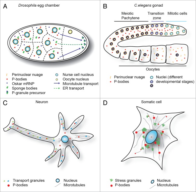
Figure 1.
mRNP granules across biology. (A) Graphic of Drosophila egg chamber indicates interaction of multiple mRNP granule types in the cytoplasm of nurse cells, and modes of transport to the germ plasm. (B) Gonad of a C.elegans hermaphrodite indicates “assembly line” maturation, from mitotic stem cells at distal end (top right) to oocytes at proximal end (bottom right). P-granules remain docked with nucleus until diplotene, then re-localize to the cytoplasm with nuclear pore components. P-bodies are distributed throughout the shared cytoplasm. (Panels A and B adapted from Voronina et al., 2011 and reproduced with permission). (C) Simplified neuronal cell schematic demonstrating specific localization of transport granules in dendrites and the axon. Interactions with P-bodies and transport along microtubules are highlighted. (D) Stressed somatic cell, indicating typical distribution of stress granules (often peri-nuclear) and P-bodies, which often dock with stress granules. Interactions of both granules with microtubules also highlighted.

