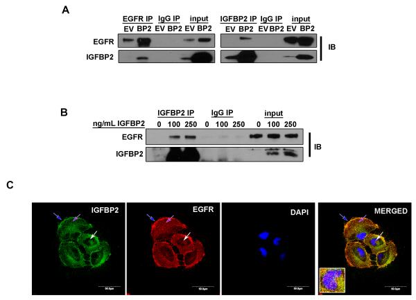
Figure 3. IGFBP2 co-precipitates and co-localizes with EGFR.
(A) Co-immunoprecipitation (IP) of IGFBP2 and EGFR in SNB19.EV control cells versus SNB19.BP2 cells analyzed by immunoblot (IB). (B) Immunoprecipitation of IGFBP2 in U87 cells starved of serum overnight then stimulated with 2 different doses of IGFBP2 for 30 minutes, analyzed by immunoblotting. (C) Confocal microscopy images of immunofluorescence staining for IGFBP2 (green), EGFR (red) and DAPI (blue) in SNB19.BP2 cells show IGFBP2 and EGFR co-localization; blue arrow = cell membrane; purple arrow = cytoplasm; white arrow = nucleus.

