Figure 2.

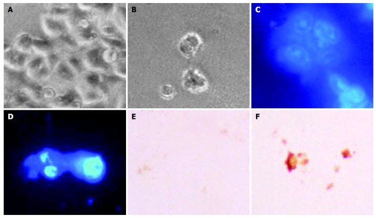
Characterization of OPC-induced cell death in SNU-C4 cells. Cells were cultured without OPC (A, C and E) or with 100 µg/mL OPC (B, D and F). Morphology (top): phase-contrast microscopy shows cell shrinkage, bubble like bleb in cell membrane and cellular detachment in OPC-treated cultures (B). SNU-C4 cells stained with DAPI staining (D). SNU-C4 cells stained with TUNEL staining (F): Scale bar, 10 µm.
